Science
University of Tokyo Unveils Advanced ‘Great Unified Microscope’
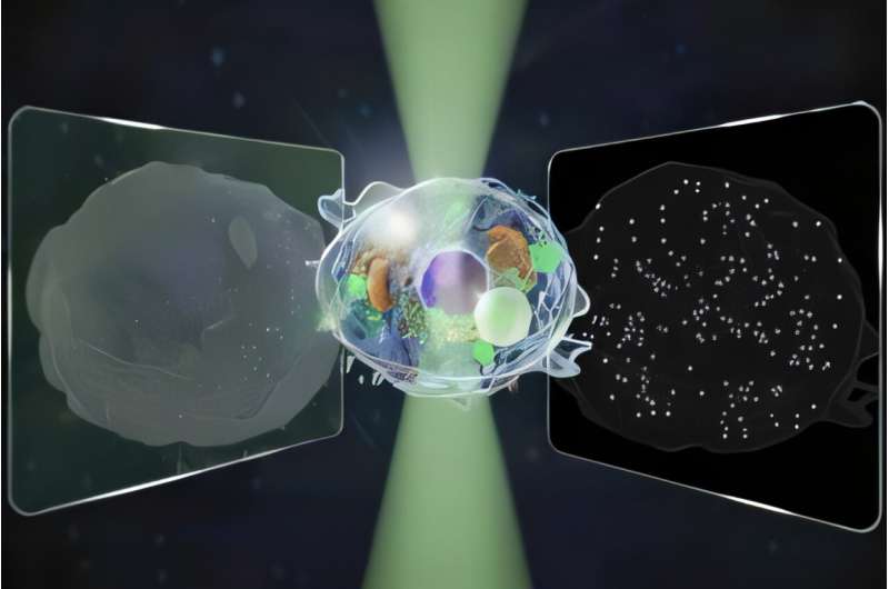
Researchers at the University of Tokyo have developed a groundbreaking microscope capable of detecting signals across a range 14 times wider than conventional models. This innovation allows for label-free observations, meaning it does not require additional dyes that can harm cells, making it suitable for long-term studies and quality control in the pharmaceutical and biotechnology sectors. The findings were published on November 14, 2025, in the journal Nature Communications.
Microscopes have been essential tools in scientific advancement since the 16th century. Yet, the demand for more sensitive and accurate equipment has led to the development of specialized techniques that often involve trade-offs. For example, quantitative phase microscopy (QPM) can detect structures over 100 nanometers but lacks the ability to observe smaller entities. This method primarily produces static images of complex cell structures.
On the other hand, interferometric scattering (iSCAT) microscopy detects minute particles, including single proteins, allowing researchers to track dynamic changes within cells. Nevertheless, it does not provide the comprehensive view that QPM offers.
Kohki Horie, a lead author on the study, expressed a desire to understand dynamic processes within living cells through non-invasive techniques. Alongside co-researchers Keiichiro Toda, Takuma Nakamura, and Takuro Ideguchi, they aimed to investigate whether simultaneous measurement of light in both directions could bridge the gap and reveal a broader range of sizes and motions in a single image.
To validate their hypothesis, the team observed cellular processes during cell death. They successfully recorded images that encoded information from both forward and backward-traveling light.
“Our biggest challenge,” Toda explained, “was cleanly separating two kinds of signals from a single image while keeping noise low and avoiding any mixing between them.” This challenge was met, allowing the researchers to quantify the motion of micro-scale cell structures alongside the movements of nano-scale particles. By analyzing both types of scattered light, they could estimate each particle’s size and refractive index, which describes how light bends or scatters when passing through various materials.
Looking ahead, Toda shared aspirations to study even smaller particles, such as exosomes and viruses, to further understand their size and refractive properties in diverse samples. The team also intends to explore how living cells undergo the process of cell death while controlling their states, cross-referencing their findings with other investigative techniques.
The development of the Great Unified Microscope signifies a significant leap forward in microscopy, offering researchers an innovative tool for studying cellular dynamics and enhancing our understanding of complex biological processes.
For more information, refer to the article titled “Bidirectional quantitative scattering microscopy” published in Nature Communications (2025). DOI: 10.1038/s41467-025-65570-w.

Springfield Central Girls Basketball Secures 57-45 Win Over Northampton

Trump Strikes Venezuela: Legal Experts Warn of Red Line Breach

Driver Charged After Fatal Christmas Day Crash Kills 2 Kids

Hornets’ Miles Bridges Exits Game with Ankle Injury, Out for Now

Lenape Surges Past Trenton 57-56 in Thrilling Semifinal Clash

Waterbury Murder Arrest: Suspect Charged After Fatal Stabbing

MAGA Fans Erupt as Fox News Host Questions Viral Fraud Claims

Exploring the Strategic Roles of the 5 Smallest US Air Force Bases

Bay Area Commuters Face Fare Increases as BART Addresses Deficit

University of Hawaiʻi at Mānoa Joins $25.6M AI Initiative for Disaster Monitoring

New Gel Offers Hope for Regrowing Tooth Enamel in Dentistry

ALMA Discovers Companion Orbiting Red Giant Star π 1 Gruis

Park Jung Min’s Endearing Moment with Hwasa Steals Show at Awards

IROS 2025 to Showcase Cutting-Edge Robotics Innovations in China
Stone Island’s Logo Worn by Extremists Sparks Brand Dilemma

Sampson County Celebrates Susie Faison’s 100th Birthday Milestone

Mary Morgan Jackson Crowned Little Miss National Peanut Festival 2025

Startup Liberate Bio Secures $31 Million for Next-Gen Therapies
-
Science2 months ago
University of Hawaiʻi at Mānoa Joins $25.6M AI Initiative for Disaster Monitoring
-
 Health2 months ago
New Gel Offers Hope for Regrowing Tooth Enamel in Dentistry
-
 Science2 months ago
ALMA Discovers Companion Orbiting Red Giant Star π 1 Gruis
-
 Lifestyle1 month ago
Park Jung Min’s Endearing Moment with Hwasa Steals Show at Awards
-
 Science3 months ago
IROS 2025 to Showcase Cutting-Edge Robotics Innovations in China
-

 Lifestyle3 months ago
Stone Island’s Logo Worn by Extremists Sparks Brand Dilemma
-
 Lifestyle2 months ago
Sampson County Celebrates Susie Faison’s 100th Birthday Milestone
-
 Lifestyle3 months ago
Mary Morgan Jackson Crowned Little Miss National Peanut Festival 2025
-
 Health3 months ago
Startup Liberate Bio Secures $31 Million for Next-Gen Therapies
-

 Health3 months ago
Top Hyaluronic Acid Serums for Radiant Skin in 2025
-

 Science3 months ago
Arizona State University Transforms Programming Education Approach
-

 Politics2 months ago
Judge Considers Dismissal of Chelsea Housing Case Citing AI Flaws