Science
AI Revolutionizes 3D Imaging of Biological Samples Using SEM
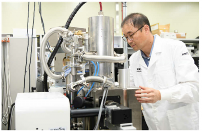
The Korea Research Institute of Standards and Science (KRISS) has unveiled a groundbreaking artificial intelligence (AI)-based image segmentation algorithm that can quickly reconstruct three-dimensional (3D) structures from two-dimensional (2D) cross-sectional images obtained through scanning electron microscopy (SEM). This innovation marks a significant advancement in the field of microscopy and biological imaging, enhancing the ability to visualize intricate cellular structures.
The development of this algorithm, led by President Lee Ho Seong and his team at KRISS, addresses a critical need in scientific research. By facilitating rapid and accurate 3D reconstruction, this technology has the potential to transform how researchers analyze biological samples. Traditional methods have often required extensive manual processing and time-consuming techniques. With this new AI approach, the process is streamlined, allowing for faster analysis and improved accuracy.
Implications for Biological Research
The ability to visualize 3D structures from SEM images opens new avenues for research across various biological disciplines. Researchers can now gain a more comprehensive understanding of cellular architecture, which is essential for studies in areas such as cell biology, pathology, and materials science. The algorithm’s capacity to automate complex imaging tasks not only enhances efficiency but also enables scientists to focus on interpreting data rather than getting bogged down by technical challenges.
In practical terms, this technology could benefit a range of applications, from studying disease mechanisms to developing new materials for medical use. For instance, researchers examining cancer cells can acquire detailed 3D models that reveal how these cells interact with their environment, leading to insights that could inform therapeutic strategies.
Future Prospects and Global Impact
As this AI-driven technology gains traction, its implications extend beyond South Korea. The KRISS initiative positions the country at the forefront of microscopy research, potentially influencing global standards and practices in biological imaging. The development aligns with a growing trend in scientific research that leverages AI and machine learning to enhance data analysis and interpretation.
The implementation of this algorithm could see widespread adoption in laboratories worldwide, fostering international collaborations and knowledge sharing. By improving the speed and precision of imaging techniques, KRISS not only contributes to the advancement of scientific research but also reinforces South Korea’s role as a leader in innovation.
In conclusion, the introduction of the AI-based image segmentation algorithm by KRISS represents a significant leap forward in the ability to reconstruct 3D structures from SEM images. This advancement promises to enhance biological research capabilities, streamline workflows, and ultimately lead to new discoveries across various scientific fields. The ongoing evolution of this technology will be closely monitored as it reshapes the landscape of microscopy and biological analysis.

Nationals’ Interim Manager Miguel Cairo Not Retained, Major Shakeup

Beloved Artist and Community Leader Gloria Rosencrants Passes Away at 94

Viking Therapeutics Unveils VK2735 Data at ObesityWeek 2025

Community Remembers Gloria Rosencrants, 94, an Artistic Spirit

Assassins Sentenced to 25 Years for Plot Against Iranian Activist

Astronomers Announce Discovery of Earth-Like Exoplanet After 20 Years

Border Patrol Chief Vows to Show Judge Violence Against ICE Agents

Urgent Update: Bodycam Footage Reveals Details in Murder Case

San Ramon Home Sells for $1.9 Million in Record-Time Deal

IROS 2025 to Showcase Cutting-Edge Robotics Innovations in China

Judge Considers Dismissal of Chelsea Housing Case Citing AI Flaws

Bravo Company Veterans Honored with Bronze Medals After 56 Years
Stone Island’s Logo Worn by Extremists Sparks Brand Dilemma

Startup Liberate Bio Secures $31 Million for Next-Gen Therapies

Indonesia Suspends 27,000 Bank Accounts in Online Gambling Crackdown

Mel Kiper Jr. Reveals Top 25 Prospects for 2026 NFL Draft

Top Hyaluronic Acid Serums for Radiant Skin in 2025

Honeywell Predicts Record Demand for Business Jets Over Next Decade
-
 Science2 weeks ago
IROS 2025 to Showcase Cutting-Edge Robotics Innovations in China
-
 Politics2 weeks ago
Judge Considers Dismissal of Chelsea Housing Case Citing AI Flaws
-
 World2 weeks ago
Bravo Company Veterans Honored with Bronze Medals After 56 Years
-

 Lifestyle2 weeks ago
Stone Island’s Logo Worn by Extremists Sparks Brand Dilemma
-
 Health2 weeks ago
Startup Liberate Bio Secures $31 Million for Next-Gen Therapies
-
 Top Stories2 weeks ago
Indonesia Suspends 27,000 Bank Accounts in Online Gambling Crackdown
-
 Sports2 weeks ago
Mel Kiper Jr. Reveals Top 25 Prospects for 2026 NFL Draft
-
 Health2 weeks ago
Top Hyaluronic Acid Serums for Radiant Skin in 2025
-
 World2 weeks ago
Honeywell Predicts Record Demand for Business Jets Over Next Decade
-

 Sports2 weeks ago
Yamamoto’s Mastery Leads Dodgers to 5-1 Victory in NLCS Game 2
-

 Politics2 weeks ago
New Jersey Voters Urged to Register Ahead of November Election
-

 Lifestyle2 weeks ago
Mary Morgan Jackson Crowned Little Miss National Peanut Festival 2025