
Health
Scientists Unveil Advanced Microscopy to Observe Flu Virus Entry

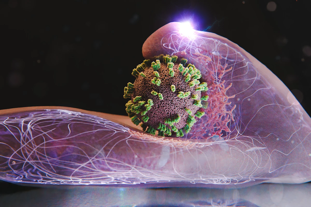
A groundbreaking study from researchers in Switzerland and Japan has developed a new microscopy technique that allows scientists to observe the influenza virus as it enters human cells. This innovative method, called virus-view dual confocal and atomic force microscopy (ViViD-AFM), combines two powerful imaging technologies to provide unprecedented real-time insights into the virus’s invasion process.
The influenza virus, notorious for its seasonal resurgence, infiltrates the human body through tiny droplets we inhale. Once inside, it uses two key proteins, hemagglutinin (HA) and neuraminidase (NA), to latch onto cells, initiating a complex process of infection. Traditional microscopy techniques have struggled to capture the rapid and minuscule movements involved in this process, leaving many aspects of the virus’s behavior poorly understood.
With the advent of ViViD-AFM, researchers can now examine living human cells in extraordinary detail. For the first time, they have visualized the intricate dance between the influenza virus and its target cells. The study revealed surprising behavior from the cells themselves, which actively engaged with the virus rather than passively allowing entry.
Yohei Yamauchi, a researcher at ETH Zurich, remarked, “The infection of our body cells is like a dance between virus and cell.” The researchers observed how individual influenza virus particles navigated the cell surface under various conditions, including scenarios where specific viral proteins were inhibited or when different strains of the virus were introduced.
The study highlighted the importance of the cell’s membrane, which changes shape during the virus’s entry. By utilizing fluorescent tags that make the virus visible, the new technique enabled scientists to track the virus without needing additional labels. This capability allows for simultaneous visualization of the viral proteins and the cellular structure, providing a comprehensive understanding of their interactions.
The findings indicated that influenza viruses require larger surface bulges on the cell, created by the protein actin, to facilitate entry. These bulges were not hindered by certain inhibitors, suggesting that the mechanism may overlap with other cellular processes. Once the virus binds to receptor clusters, it triggers signals that prompt the cell to envelop the virus in a clathrin coat, ultimately pulling the virus inward and encapsulating it in a vesicle for transport to the cell nucleus.
The implications of this advanced microscopy technique extend beyond just studying influenza. Researchers anticipate that ViViD-AFM could become a vital tool in evaluating the efficacy of antiviral medications in real time, as well as examining the behavior of various viruses and the interactions of vaccines with human cells.
This breakthrough offers a new “window” into cellular activity, potentially reshaping our understanding of how viruses operate and how medicines can be developed to combat them. The research findings have been published in the journal PNAS, marking a significant step forward in virology and drug research.

Robinhood Stock Surges 2.5% Today Amid Tech Market Recovery

Philadelphia’s Earth Month Kicks Off with Free Events in Parks

GSK’s Inhaler Decision Sparks Health Concerns for Children

Bradford’s Fish Fry Fridays Offer Community Spirit and Flavor

Speculation Rises on Shutdowns During Trump’s Potential Presidency

Samsung Galaxy Watch 9 to Feature Larger Battery, Report Reveals

Inventors Find New Purpose Through Unconventional Paths

UConn Huskies Aim for Back-to-Back Titles in 2026 NCAA Tournament

U.S. Mine Countermeasure Ships Dock in Malaysia for Logistics Stop

University of Hawaiʻi at Mānoa Joins $25.6M AI Initiative for Disaster Monitoring

$2.2 Million Boost for Cancer Research and Training in Hawaiʻi

ALMA Discovers Companion Orbiting Red Giant Star π 1 Gruis

Sacituzumab Govitecan Shows Promise for HR+/HER2− Breast Cancer

币安人生 Sees $15.92 Million in Trading Volume Amid Market Fluctuations

New Gel Offers Hope for Regrowing Tooth Enamel in Dentistry

CAVA Group Shares Surge 5.1% Amid Mixed Analyst Ratings

Christian Bale in Talks to Join Leonardo DiCaprio in Heat 2

Supreme Court to Review Birthright Citizenship for Children of Immigrants
-
Science9 months ago
University of Hawaiʻi at Mānoa Joins $25.6M AI Initiative for Disaster Monitoring
-
 Health8 months ago
$2.2 Million Boost for Cancer Research and Training in Hawaiʻi
-
 Science9 months ago
ALMA Discovers Companion Orbiting Red Giant Star π 1 Gruis
-
 Health7 months ago
Sacituzumab Govitecan Shows Promise for HR+/HER2− Breast Cancer
-
Politics5 months ago
币安人生 Sees $15.92 Million in Trading Volume Amid Market Fluctuations
-
 Health9 months ago
New Gel Offers Hope for Regrowing Tooth Enamel in Dentistry
-
 Business8 months ago
CAVA Group Shares Surge 5.1% Amid Mixed Analyst Ratings
-
 Entertainment9 months ago
Christian Bale in Talks to Join Leonardo DiCaprio in Heat 2
-
 Politics8 months ago
Supreme Court to Review Birthright Citizenship for Children of Immigrants
-

 Lifestyle9 months ago
Art Deco and Traditional Designs Set to Transform Homes in 2026
-

 Lifestyle8 months ago
Fernando Mendoza Secures Maxwell and O’Brien Awards Ahead of Heisman
-

 World8 months ago
Swiss Residents Reject 50% Inheritance Tax on Wealthy